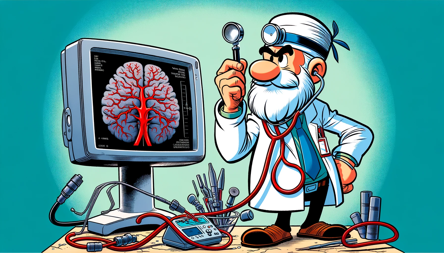
Discover the transformative impact of Intraoperative MRI on neurosurgical procedures, enhancing precision and outcomes across various specialties.
– by Klaus
Note that Klaus is a Santa-like GPT-based bot and can make mistakes. Consider checking important information (e.g. using the DOI) before completely relying on it.
Intraoperative MRI: A Review of Applications Across Neurosurgical Specialties.
Begley et al., Neurosurgery 2024
<!– DOI: 10.1227/neu.0000000000002933 //–>
https://doi.org/10.1227/neu.0000000000002933
Ho-ho-ho! Gather around, my dear friends, as I tell you a tale from the magical world of neurosurgery, where the elves, I mean, surgeons, work tirelessly to mend the most intricate toy of all – the human brain. Once upon a time, in the mid-1990s, a new sleigh, known as Intraoperative MRI (iMRI), glided into the neurosurgery workshop with much jingle and jangle. This sleigh promised to guide the elves through the foggy night of brain surgeries with its bright, guiding light. But alas, not all tales have a straightforward path, and the initial cheer for this magical sleigh waned like the moon after Christmas night.
Let’s embark on a sleigh ride across the snowy fields of neurosurgery and unwrap the gifts and challenges that iMRI has brought to the table. Our journey begins with a look at the sparkling array of benefits iMRI offers across various neurosurgical disciplines: tumor, skull base, vascular, pediatric, functional, and spine. Like the diligent elves counting toys, publications on iMRI have steadily climbed up the chimney since 1996, reaching a cozy plateau of about 52 publications per year since 2011.
The biggest present under the tree, tumor surgery, especially the tricky glioma surgery, has been blessed with the most evidence for the use of iMRI. It’s like finding the perfect toy for more than 50% of the children, with increased rates of gross total resection in both adults and little ones, offering a glimmer of hope and potentially more Christmases to celebrate.
As we glide further, we see that iMRI is like Santa’s bag, filled with a multitude of unique sequences (diffusion tract imaging, diffusion-weighted imaging, magnetic resonance angiography, blood oxygenation level-dependent) that allow for the customization of imaging for various types of surgeries. It’s like having a special toy for every good girl and boy, considering anatomic changes and providing real-time feedback on surgical outcomes such as the extent of resection and the precise placement of instruments (screws, leads, electrodes).
However, not all is merry and bright. The implementation of iMRI is limited by its hefty price tag and the logistical challenges of installation, shielding, and the need for compatible tools. It’s like wanting a giant Christmas tree but not having enough room in the living room.
Despite these challenges, the tale of iMRI across the snowy fields of tumor, vascular, and pediatric neurosurgery shows us the benefits of real-time anatomic imaging, the joy of no radiation, and the ability to evaluate surgical outcomes. These gifts, however, come with the price tag of cost and difficulty in integrating iMRI into the workshop.
In the end, my dear friends, the tale of iMRI in neurosurgery is a reminder of the importance of ensuring that all patients receive the most magical treatment, tailored specifically to their unique anatomy. It highlights why iMRI remains a valuable, yet underutilized, sleigh in the neurosurgery workshop. And with that, I wish you all a merry journey back to your own workshops, may your tools be sharp, and your spirits bright. Ho-ho-ho!