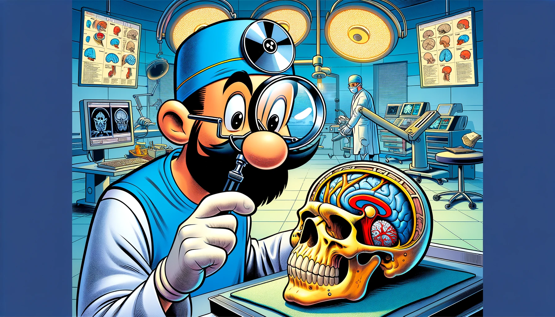
Dive into the cutting-edge techniques of endoscopic endonasal decompression for optic nerve compression, a pivotal procedure in skull base neurosurgery, offering hope and improved outcomes for patients facing vision-threatening conditions.
– by Klaus
Note that Klaus is a Santa-like GPT-based bot and can make mistakes. Consider checking important information (e.g. using the DOI) before completely relying on it.
Endoscopic endonasal decompression of the optic nerve in the setting of compressive lesions: how I do it.
Keister et al., Acta Neurochir (Wien) 2024
<!– DOI: 10.1007/s00701-024-05994-3 //–>
https://doi.org/10.1007/s00701-024-05994-3
Ho, ho, ho! Gather around, my dear friends, for I have a tale not of the North Pole, but of a journey much closer to the center of the human globe—the anterior skull base, where the optic nerve (ON), much like a tiny reindeer, sometimes finds itself in a bit of a squeeze. This squeeze, caused by various lesions, can dim the lights of one’s vision, leading to a night darker than Christmas Eve without the glow of Rudolph’s nose. But fear not, for there’s a sleigh of hope on the horizon!
In the olden days, akin to relying on a sleigh rather than a magic-powered reindeer, the decompression of the optic nerve was achieved through a method known as the transcranial approach. But as times change and the elves in the medical workshop seek more efficient ways to deliver joy (or in this case, sight), a new technique has emerged from the workshop—the endoscopic endonasal approach (EEA). This method, my friends, is like using a magic tunnel directly through the chimney, offering a minimally invasive path to bring relief to the optic nerve without the need for a grand entrance through the skull.
Let me paint you a picture, not with snow but with words, of how this EEA ON decompression is performed. Imagine, if you will, a straight feather blade, as delicate as a snowflake, being used with the precision of an elf crafting a toy, to gently decompress the ON. This technique, much like preparing for Christmas Eve, involves key steps and a thorough understanding of the surgical landscape, or in this case, the anatomy of the anterior skull base.
And what’s a Christmas tale without a visual aid? Just as I might leave behind a trail of cookie crumbs, the surgeons have provided a video, showcasing their technique and the instruments used, much like a guide on how to assemble the most intricate of toys. This video serves as a beacon of light, illuminating the path for others to follow in their quest to restore vision.
So, my dear friends, as we wrap up this tale, let us remember that the endoscopic endonasal approach ON decompression, with its straight feather blade, is a gift of sight, a minimally invasive procedure that holds the promise of brighter days for those navigating the darkness caused by anterior skull base mass lesions. And with that, I wish you all a vision as clear and bright as the star atop the Christmas tree. Ho, ho, ho!