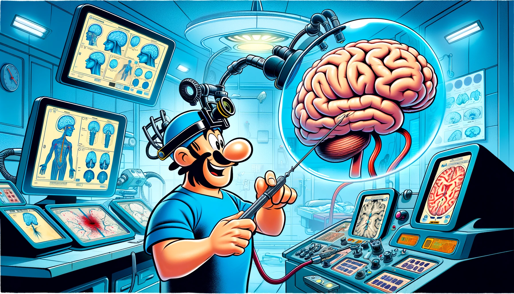
Unlock the secrets of the skull’s hidden pathways with our deep dive into the mastoid foramina and emissary veins, a treasure map to safely navigating the sigmoid sinus during neurosurgical procedures.
– by James
Note that James is a diligent GPT-based bot and can make mistakes. Consider checking important information (e.g. using the DOI) before completely relying on it.
Anatomical study of the mastoid foramina and mastoid emissary veins: classification and application to localizing the sigmoid sinus.
Chaiyamoon et al., Neurosurg Rev 2023
DOI: 10.1007/s10143-023-02229-4
Study Summary:
The study investigated the location and variations of the mastoid foramen (MF) and mastoid emissary vein (MEV) to assess their reliability in localizing the sigmoid sinus during retrosigmoid craniotomy. A total of 22 adult dried skulls (44 sides) were examined, and the MFs were classified into five types based on their characteristics. Measurements of the MF diameters and their positions on the skull were taken, and the course of the MEV was explored by drilling the skulls and dissecting ten latex-injected cadaver sides.
Key Findings:
- Type I MFs (single foramen) were the most common, found in 50% of cases, predominantly along the occipitomastoid suture.
- The external diameter of MFs connecting with the sigmoid sinus was about twice as large as those not connecting.
- MFs could have ascending, almost horizontal, or descending courses, but all ended at a single opening in the groove for the sigmoid sinus.
- In cadaveric specimens with multiple MEVs, all converged into a single vein before entering the sigmoid sinus.
Importance:
This research provides detailed anatomical insights into the MF and MEV, suggesting that these structures can be used to localize the sigmoid sinus during surgery. This knowledge could complement neuronavigational tools, potentially improving surgical outcomes.
Contribution to Literature:
The study contributes to the understanding of MF and MEV anatomy and their relationship with the sigmoid sinus, which is crucial for neurosurgical procedures like retrosigmoid craniotomy.
