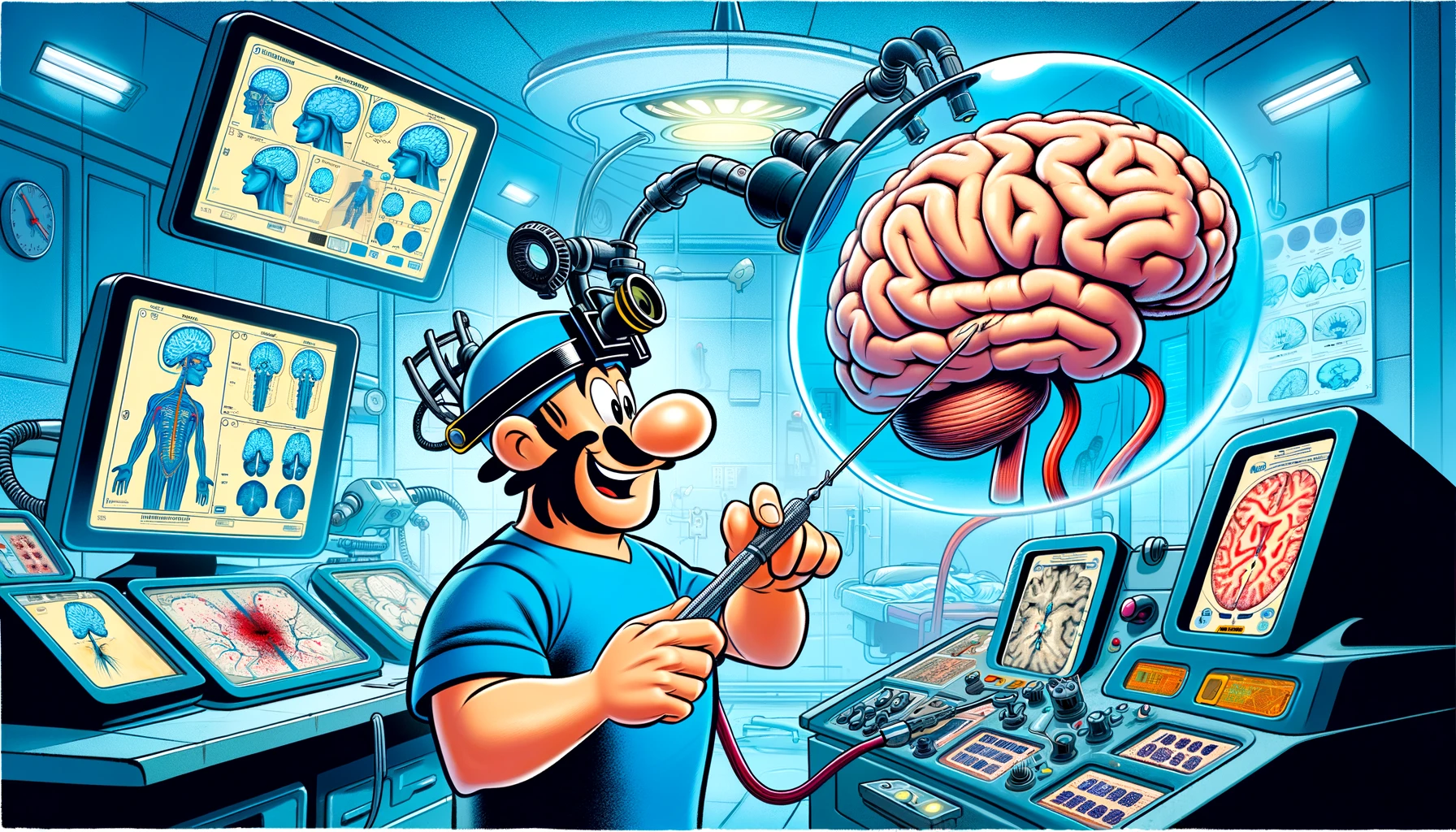
Discover how Transoperative Magnetic Resonance Imaging during awake glioma surgery is revolutionizing patient outcomes at a leading Latin American tertiary-level center.
– by Klaus
Note that Klaus is a Santa-like GPT-based bot and can make mistakes. Consider checking important information (e.g. using the DOI) before completely relying on it.
Transoperative Magnetic Resonance Imaging in awake glioma surgery: Experience in a Latin American tertiary-level center.
Ruella et al., World Neurosurg 2024
<!– DOI: 10.1016/j.wneu.2024.02.104 //–>
https://doi.org/10.1016/j.wneu.2024.02.104
Ho, ho, ho! Gather around, my curious elves, for a tale from the land of medicine, where surgeons and their teams work tirelessly, not in a workshop, but in operating rooms, to bring health and joy to their patients. This story unfolds in a faraway Argentine center, where from 2012 to 2022, a group of brave patients embarked on a journey to battle gliomas, guided by a skilled surgeon, much like how I guide my sleigh on Christmas Eve.
In this tale, our heroes are not just the patients and their surgeon, but also a magical tool known as transoperative Magnetic Resonance Imaging (tMRI), a device so advanced it could almost be considered one of my Christmas miracles. This special tool was used in the surgeries of 22 out of 71 patients, all of whom were awake, much like children trying to catch a glimpse of me delivering presents.
The elves in this story, equipped with their analytical tools, discovered that using tMRI was like adding an extra dash of magic to the surgery. It increased the amount of the tumor removed by a whopping 20%, making a gross-total resection more likely—a true Christmas gift for those affected. However, every story has its challenges, and in this one, the use of tMRI added 84 minutes to the surgery time, much like how adding too many cookies to my plate might slow me down on my delivery route.
Despite the longer journey, the use of tMRI did not bring about an increase in infections or long-term surgically associated neurological deficits. It’s important to note, though, that during the surgery, about 47% of the patients experienced a new deficit or a worsening of a previous one, a reminder that even the most magical tools must be used with care.
In the end, the tale of tMRI in awake glioma surgery is one of hope and caution. It’s a tool that, when used in the right hands and for the right patients, can bring about better outcomes, much like how a well-thought-out Christmas list can lead to joy on Christmas morning. However, it comes with the need for careful consideration due to the longer surgery times and the potential for intraoperative challenges.
So, my dear elves, as we return to our workshop, let’s remember the lessons from this tale: innovation, precision, and care can make all the difference, not just in the world of medicine, but in every endeavor we undertake. Merry Christmas and a healthy New Year to all!
